Posted April 16, 2024
Hamilton and Bowdoin Colleges’ Women’s Lacrosse Teams Dedicate Game to Fund Alzheimer's Disease Research
Cure Alzheimer’s Fund is honored to be the recipient of donations raised by the Hamilton and Bowdoin Colleges’ women’s lacrosse teams.
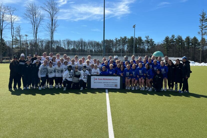
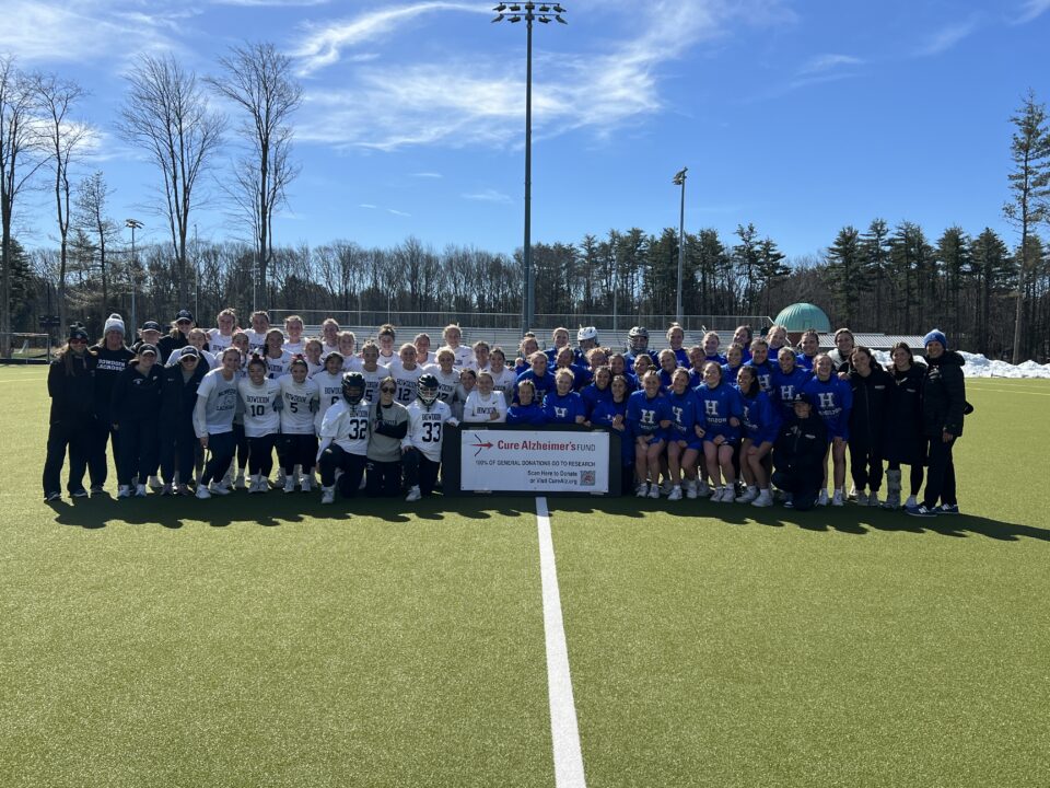
The Continentals from Hamilton College @hamiltonwomenslacrosse in Clinton, New York, were hosted by NESCAC rival Polar Bears from Bowdoin College @bowdoinwlax in Brunswick, Maine on Saturday, March 30. The teams joined forces to dedicate their game to raising awareness and contributions for research in honor of those who have been impacted by Alzheimer’s disease. More than $900 was raised for research.
“Over the course of their lives, women are more likely to be both caregivers and patients impacted by Alzheimer’s disease,” said Meg Smith, CEO of Cure Alzheimer’s Fund. “These young women inspire us by educating their communities about this relentless disease and our work to end it, and by taking control of their futures and reducing their own risk of dementia through education and exercise.”
There are 6.9 million people in the United States—and 50 million people throughout the world—currently living with an Alzheimer’s diagnosis, and experts estimate there may be as many as three times more people living with the disease who have not yet been diagnosed. Both men and women develop Alzheimer’s disease, but two-thirds of all patients are women, and women are frequently the primary caregivers for spouses, partners, parents and others with the disease.
“I am grateful to have partnered with my friend, Liz Grote, and her Bowdoin Lacrosse team,” said Patty Kloidt, coach of the Hamilton Women’s Lacrosse team. “She and I, along with members of our teams have experienced loved ones who suffered from this disease. We want our communities to be aware of the Cure Alzheimer’s Fund and consider supporting their amazing work. Too many people, including caregivers, are suffering from this horrible disease. Please spread the word about the Cure Alzheimer’s Fund and join in the fight for our future generations.”